Friday, July 27, 2012

Monday, July 23, 2012
PET (F18FDG) - Atherosclerosis

Clinical history: multiple myeloma and temporal arteritis (inflammation and dammage to the blood vessels that supply the head, particularly the large or medium arteries that branch from the neck and supply the temporal area), weight loss.
PET scan for temporal arteritis can produce detailed images of the blood vessels and highlight areas of inflammation. PET imaging for atherosclerosis of carotid and peripheral arteries have been documented in several studies see below.
The patient was injected with 15 mCi F18 FDG and imaged from brow to mid-thigh at 1.5 hours post injection, with CT for attenuation correction. blood glucose at the time of injection was 90 mg/dl.
Findings - The images demonstrate diffuse metabolic activity in the carotid arteries suggesting active atherosclerosis or inflammation. Diffuse activity is present in the aorta, large vessels of the thorax, pelvis, upper and lower extremities...
There was no evidence of metabolically active neoplasm.
Further reading
Friday, July 20, 2012
July 20, 2012 UMDNJ Newark
Welcome to the Advisory Committee Meeting!!
Agenda
Update on Current Students
Review of Applicant Pool for 2013 Program
Update on Competencies and Required Program Content as per Accrediting Bodies
Documentation of Student Performance at Clinical Sites - New Evaluation Form
Program Status and Accreditation
Updates from Affiliates
Open Discussion
Thursday, July 19, 2012
Cutaneous Metastases
Breast cancer is known to metastasize
to anywhere in the body, either by hematogenous or lymphatogenous
routes. Cancers that have the highest tendency to metastasize to the
skin include melanoma, breast, nasal sinuses, larynx and oral cavity.

 Another example of a female with a clinical history of breast cancer who presents with "nodular" cutaneous metastases. image includes PET WB coronal slice and comparative transverse PET and CT slices. Localized nodules are the most common presentation, occurring in 10% of patients.
Reading:
Cutaneous metastases of breast carcinoma: a case report.
Cutaneous and subcutaneous metastases of adenocarcicoma of the colon and rectum
Excluding melanoma, cutaneous
metastases occur more often in breast cancer than in other diseases
in women and is the most often encountered in clinical practice. In
a recent study, that included 12146 patients with internal
malignancies, the rate of CM associated with breast carcinoma was
2.42 percent.

PETCT - subcutaneous metastases - transverse image shows extent of disease located in the soft tissue at the upper lobe of the liver.
projection image

Reading:
Cutaneous metastases of breast carcinoma: a case report.
Cutaneous and subcutaneous metastases of adenocarcicoma of the colon and rectum
Wednesday, July 18, 2012
Lung Scan - renal uptake

Technique: Patient received 20 mCi Xe133 ventilation, followed by 30 mCi Tc99m MAA for lung perfusion imaging.
There was renal uptake noted on the perfusion images which was an unexpected finding. Following investigation it was found that the patient had come into the medical center the previous night through the emergency room and a lung scan had been started. Due to equipment malfunction the perfusion portion of the scan was not completed, and was to be repeated the following morning. The uptake by the kidneys was determined to be most likely due to biological breakdown of the Tc99 MAA from the previous night, which was being removed from the body through the kidneys.
Thanks to Cheryl Casale, 2004.
Monday, July 16, 2012
Perfusion Lung Scan - Artifact
Perfusion lung scan - Tc99m MAA
Images obtained on two consecutive patients demonstrated mild uptake in the salivary glands, thyroid and stomach. This represents free technetium due to either poor kit preparation or kit breakdown. According to the package insert no less than 90% of the pertechnetate added to a reaction vial is bound to aggregate at preparation time and remains bound throughout the 6-8 hour lifetime of the preparation (depending on the manufacturer).
Use of Tc MAA after 8 hours of kit preparation has been shown to have an increase in agglomeration. It is recommended for preparation and use of Tc 99m MAA, that a fresh dose should be used for imaging.


Friday, July 13, 2012
Extravasation
Extravasation is the accidental administration of intravenously infused medications into the extravascular space around the infusion site, either by leakage due to brittle veins, previous venipuncture or mispositioned needles or IV catheters.
Infiltration, the result of an extravasation, is the diffusion or accumulation of substance not normal to it in amounts in excess of the norm.
IV infusion should be free flowing. The arm with the infusion should not begin to swell, get red (erythemia), get hot and the patient should not notice any pain or burning. It is best to assure good access with saline prior to injecting radioactive materials. Should you notice any problems with the injection stop immediately. Keep in mind that often the volume of radiopharmacuetical dose are small and a dose may be infiltrated before you can react.
With scans such as a bone scan, the extravasation of a dose may not be critical to the scan. However the extravasation of labeled white blood cells, PET doses, any study requiring dynamic acquisition such as renal scans may result in the patient having to return for a repeat study. Take care when injecting.
 Note the increased uptake of radiopharmaceutical (TC MDP) in the left hand, site of injection.


Infiltrated dose of TcMDP on this bone scan shows the radiopharmaceutical tracking up the left arm with a visible lymph node in the left axilla. Not all injections are going to be perfect. If the dose is infiltrated it can be covered with lead to attenuate the accumulated dose or the arm can be moved from the image. This will provide better count statistics from the image.
Infiltration, the result of an extravasation, is the diffusion or accumulation of substance not normal to it in amounts in excess of the norm.
IV infusion should be free flowing. The arm with the infusion should not begin to swell, get red (erythemia), get hot and the patient should not notice any pain or burning. It is best to assure good access with saline prior to injecting radioactive materials. Should you notice any problems with the injection stop immediately. Keep in mind that often the volume of radiopharmacuetical dose are small and a dose may be infiltrated before you can react.
With scans such as a bone scan, the extravasation of a dose may not be critical to the scan. However the extravasation of labeled white blood cells, PET doses, any study requiring dynamic acquisition such as renal scans may result in the patient having to return for a repeat study. Take care when injecting.
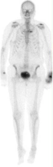

Additional views of the hand in the palmar and lateral views demonstrates increased uptake in the tissue of the left hand.

PET scan with extravasation of F18FDG in the right upper arm. In the case of PET imaging the infiltration of the dose results in invalid standard uptake value (SUV) which is determined by the exact patient dose (Pre-calibration - post-calibration) and the patient weight. If the patient dose is made inaccurate by the infiltrated dose the SUV becomes invlaid. The scan however may still be diagnostic if enough of the radiopharmaceutical has been properly delivered. The small dot of activity in the right shoulder may represent a lymph node due to the infiltration.
This patient was re-scanned with the area of extravasation out of the field of view and determined to be of diagnostic value.

Subscribe to:
Posts (Atom)