Saw this as part of a patient care presentation at work. A great video, from the Cleveland Clinic, to attempt understand everything that evolves around us each day at work and in life in general...
Showing posts with label Patient care. Show all posts
Showing posts with label Patient care. Show all posts
Wednesday, May 28, 2014
Empathy: The Human Connection to Patient Care
Saw this as part of a patient care presentation at work. A great video, from the Cleveland Clinic, to attempt understand everything that evolves around us each day at work and in life in general...

Thursday, September 6, 2012
Patient orientation
We strive to provide the best quality images possible. However, patients do not always co-operate due to severe pain, which can make lying still on an imaging table very difficult. As technologist it is our job to get the best possible images while accommodating the patient. this may mean making adjustments to the department protocol. If a whole body sweep is part of the protocol, this may mean imaging spot shots instead, or reducing time per image where acceptable.
One alternative to whole body imaging for a patient who can not lie on their back may be to have the patient lie on their stomach or on their side. Patients who are claustrophobic may be able to manage a whole body scan on their stomach better then if they are on their backs. If the patient position must be adjusted for patient comfort make sure the camera orientation is changed accordingly.
 The above template is from a Siemens PETCT scanner. The acquisition page provides four boxes to the right of the upright figure, with small images for patient positioning (supine, prone, right side and left side). The last image of the left side position is depressed, therefore the correlating image on the bottom half of the page demonstrates the patient position. Before acquiring the image ensure the patient is in the same orientation.
 Above is an example of the topogram from a PET scan patient with a history of lung cancer that had difficulty breathing while lying on her back. By having the patient lie on her side (her choice for comfort) she was able to manage the procedure without difficulty.
Quality imaging does mean patient comfort.
One alternative to whole body imaging for a patient who can not lie on their back may be to have the patient lie on their stomach or on their side. Patients who are claustrophobic may be able to manage a whole body scan on their stomach better then if they are on their backs. If the patient position must be adjusted for patient comfort make sure the camera orientation is changed accordingly.


Quality imaging does mean patient comfort.
Friday, July 13, 2012
Extravasation
Extravasation is the accidental administration of intravenously infused medications into the extravascular space around the infusion site, either by leakage due to brittle veins, previous venipuncture or mispositioned needles or IV catheters.
Infiltration, the result of an extravasation, is the diffusion or accumulation of substance not normal to it in amounts in excess of the norm.
IV infusion should be free flowing. The arm with the infusion should not begin to swell, get red (erythemia), get hot and the patient should not notice any pain or burning. It is best to assure good access with saline prior to injecting radioactive materials. Should you notice any problems with the injection stop immediately. Keep in mind that often the volume of radiopharmacuetical dose are small and a dose may be infiltrated before you can react.
With scans such as a bone scan, the extravasation of a dose may not be critical to the scan. However the extravasation of labeled white blood cells, PET doses, any study requiring dynamic acquisition such as renal scans may result in the patient having to return for a repeat study. Take care when injecting.
 Note the increased uptake of radiopharmaceutical (TC MDP) in the left hand, site of injection.


Infiltrated dose of TcMDP on this bone scan shows the radiopharmaceutical tracking up the left arm with a visible lymph node in the left axilla. Not all injections are going to be perfect. If the dose is infiltrated it can be covered with lead to attenuate the accumulated dose or the arm can be moved from the image. This will provide better count statistics from the image.
Infiltration, the result of an extravasation, is the diffusion or accumulation of substance not normal to it in amounts in excess of the norm.
IV infusion should be free flowing. The arm with the infusion should not begin to swell, get red (erythemia), get hot and the patient should not notice any pain or burning. It is best to assure good access with saline prior to injecting radioactive materials. Should you notice any problems with the injection stop immediately. Keep in mind that often the volume of radiopharmacuetical dose are small and a dose may be infiltrated before you can react.
With scans such as a bone scan, the extravasation of a dose may not be critical to the scan. However the extravasation of labeled white blood cells, PET doses, any study requiring dynamic acquisition such as renal scans may result in the patient having to return for a repeat study. Take care when injecting.
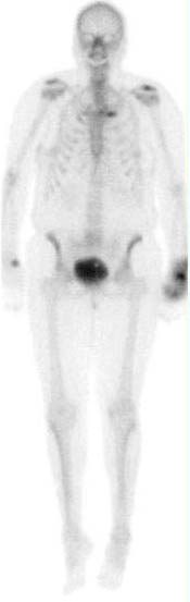

Additional views of the hand in the palmar and lateral views demonstrates increased uptake in the tissue of the left hand.

PET scan with extravasation of F18FDG in the right upper arm. In the case of PET imaging the infiltration of the dose results in invalid standard uptake value (SUV) which is determined by the exact patient dose (Pre-calibration - post-calibration) and the patient weight. If the patient dose is made inaccurate by the infiltrated dose the SUV becomes invlaid. The scan however may still be diagnostic if enough of the radiopharmaceutical has been properly delivered. The small dot of activity in the right shoulder may represent a lymph node due to the infiltration.
This patient was re-scanned with the area of extravasation out of the field of view and determined to be of diagnostic value.

Subscribe to:
Posts (Atom)